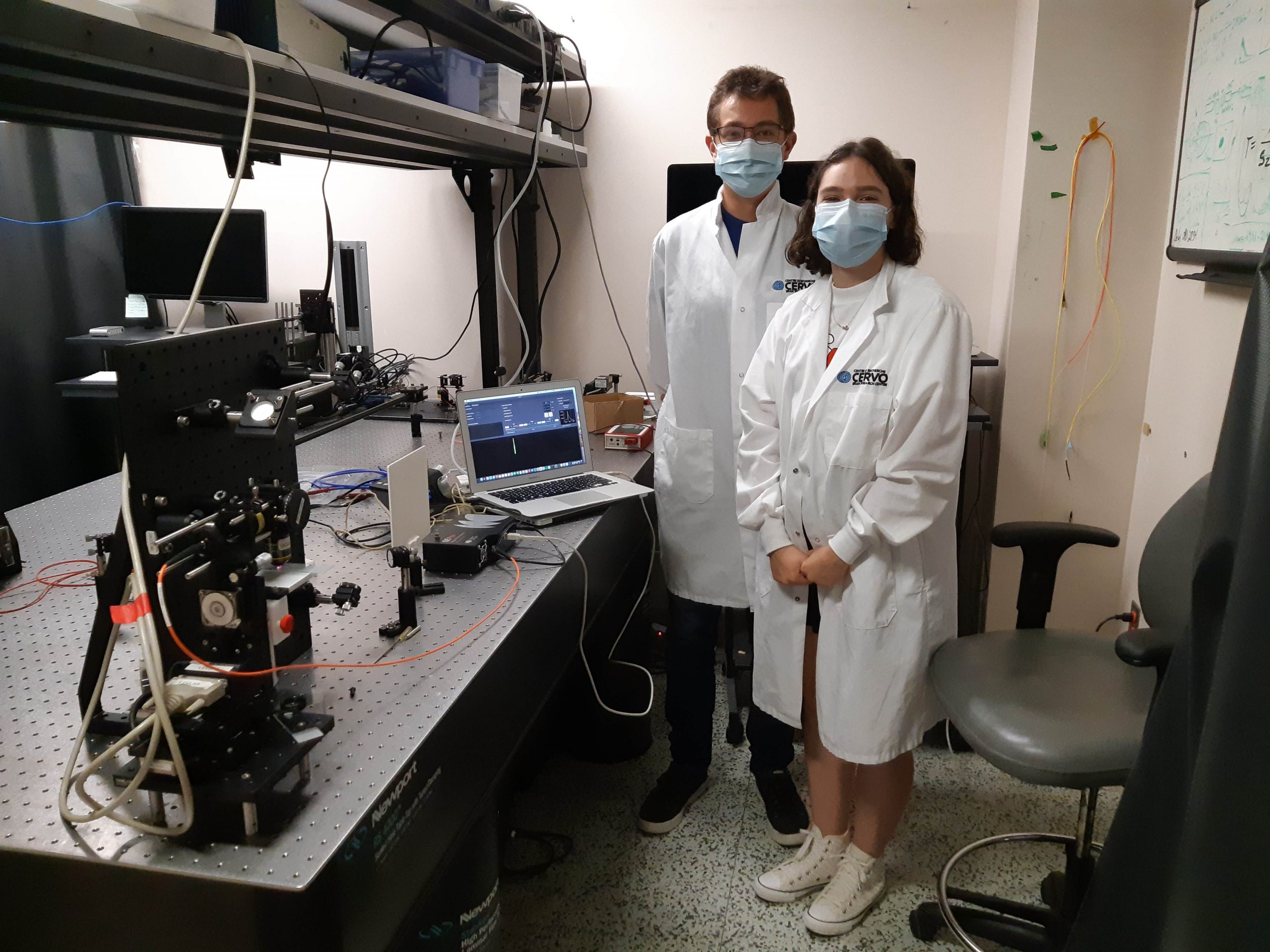

Erik Bélanger is hard at work with Cyril Bories on a project to characterize the limits of our myelin imaging technique (CARS). With a new animal model of local demyelination, samples of the corpus callosum (the highly myelinated part of the brain that connects the two hemispheres) or the striatum are imaged and the myelin thickness will be characterized.